Advanced Dental Technology La Crosse
Comfortable, Same-Day Care Offered with the Latest Technology
At All Smiles Implants and Family Dental, we want all of our patients to enjoy visiting our office, and we take important steps to make that happen. Advanced dental technology in La Crosse, like our digital impression system, cone beam CT scanner, and intraoral cameras, allow us to offer a number of same-day treatments for your convenience and comfort. You can learn more about these cutting-edge instruments by reading below, and we want you to know that we’re only a phone call away when your smile needs attention!
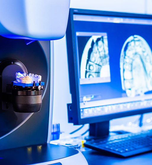
Digital Dental Impressions
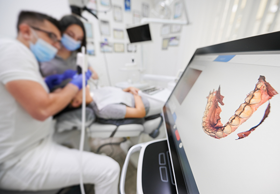
In some offices, dental impressions are taken using quick-hardening putty, which can not only induce gagging, but can sometimes require multiple tries before getting a perfect impression. At All Smiles Implants and Family Dental, we’ve invested in precision and comfort during this process by utilizing our state-of-the-art digital impression system. By simply waving a wand around your mouth, our team will be able to create a 3D digital blueprint of your gums and dental structure which we can then send off to our in-house milling machine or off-site dental laboratory.
CT/Cone Beam Scanner
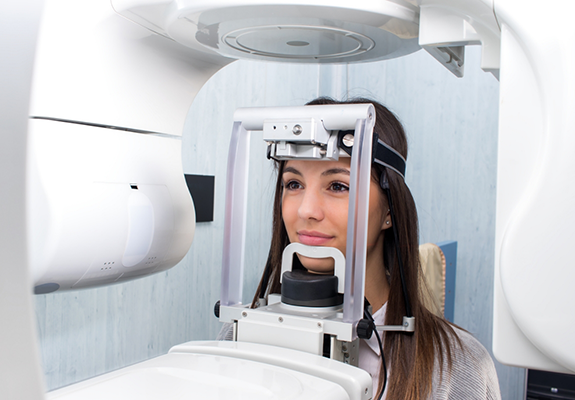
When it comes to procedures like dental implant placement and root canal treatment, having a detailed roadmap of the mouth is critical in order to achieve the best results possible. That’s why we capture cone beam CT scans of the mouth before the procedure, allowing Dr. Saphner to plan each step in detail. This device rotates around the head and puts thousands of photos together to create a 3D blueprint of the entire face. It allows Dr. Saphner to see the location of the sinuses, jawbone density, tooth roots, and even the facial nerves.
Intraoral Cameras
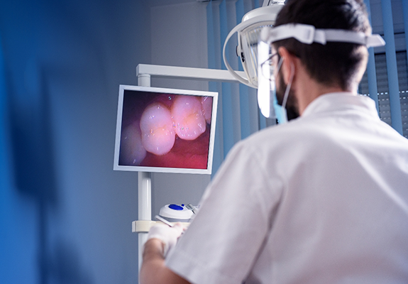
At your routine dental checkups, you may see us use our intraoral camera to gain a better view of your mouth. This device is shaped like a small wand and has a camera at the tip of it, allowing our team to capture video footage and still images of hard-to-reach areas within the mouth. We utilize these images to provide our patients with a visual educational resource when diagnosing issues and going through our treatment recommendations, and for ourselves to keep track of teeth that we’re monitoring for decay.